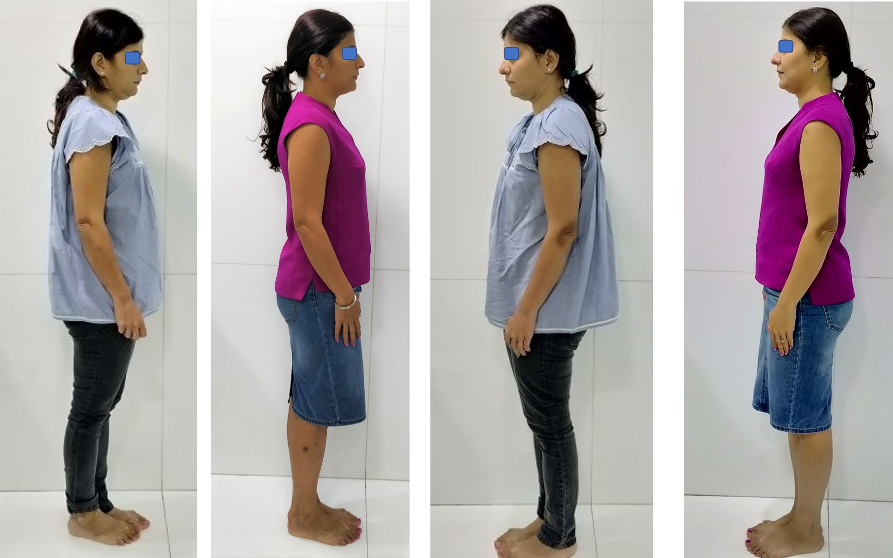
Occlusal correction and restorative treatment resulted in a remarkable improvement in the posture of the patient, pertaining to the head position and curvature of the spine. (Photograph: by Dr. Rinku Jain)
The relationship between stomatognathic and postural system has been investigated by
many health care professionals. Posture and the mandibular function are strongly
influenced by the position of the teeth. We know any missing tooth if not replaced for
long period of time may lead to change in the bite, because of the change in the position
of the adjacent and/ or opposing teeth, especially in pateints with poor oral hygiene. A
successful clinical outcome of any dental restorative work depends on proper occlusal
harmony. With advanced digitization, we can measure dynamic occlusal forces and
achieve occlusal stability with T-Scan. Here is a case report with treatment of traumatic
bite and its influence on posture, before and after achieving the occlusal harmony using
T-Scan.
Introduction:
“The relation between dental occlusion, body posture and temporomandibular disorder is a controversial topic in dentistry, though the role of dental occlusion in the development of TMDs cannot be overruled”(2). “During the routine oral examination, the signs and symptoms of dental occlusal disease must be noted, and the patient
should be educated about the further diagnosis and treatment” (2). This is a case report showing how occlusal changes can lead to postural changes. This case report details the correction of traumatic bite with restorative work and measurement of occlusal forces with T-Scan. It shows the importance of correction of the traumatic bite at the earliest to prevent TMD and postural changes. It was observed that minimal changes in occlusion can affect the posture.
Case report:
Patient aged 38, reported to our office to get her missing teeth fixed. She informed of habitual chewing of food only on the right side of the jaw.
Medical History:
Chronic sinusitis & headaches 2-3 times in a week.
No other relevant medical history.
Dental History:
Extraction of upper left molar a year back and extraction of a lower right
molar 2 years back.
Intra oral examination:
Intraoral photographs.
Incisal attrition with 12, 11, 21, 22, 31, 32, 41, 42, 43 (Fig 1, 2, 3, 4)
Missing tooth no 26 & 46
Reduced mesio-distal space between 45 & 47 (Fig 3)
Mesialy tilted 47 (Fig 5), Supra erupted 36 (Fig 6),
Cervical abrasion with 23, 34, 44
Caries with 11, 12, 16, 17, 21, 22, 24, 25, 27, 45, 47, 48
Extraoral examination:
No relevant signs related to TMJ, and other facial structures
Investigations: CBCT.
Postural photographs.
Fig 1: incisal attrition and proximal caries with upper anterior teeth
Fig 2: incisal attrition and proximal caries with upper anterior teeth
Fig 3: reduced edentulous space between 47 & 45
Fig 4: incisal wear of 41, 42,& 43
Fig 5: mesially tilted and rotated 47
Fig 6: supraerupted 36
Treatment plan:
Oral prophylaxis followed by treatment of carious teeth with biomimetic restorations. An incisal build-up for all the anterior teeth with stress-reduced direct composite restoration, Indirect Sinus Lift & implant placement with 26. Occlusal force analysis with T-Scan.
Discussion:
The patient had fair oral hygiene. The extracted teeth were not replaced hence there was supra eruption of the opposing molar on the left side and mesial inclination of 47, leading to traumatic bite, thus leading to incisal attrition of upper and lower anterior teeth and abrasion with 23, 34 & 44. Pre-treatment and post-treatment postural photographs were taken to see if the correction of occlusion had any effect on posture.
After explaining to the patient, about the intraoral findings and the importance of occlusal correction not only for the prognosis of the implant but also for the health of the teeth and mouth, the patient agreed to the planned treatment.
Firstly oral prophylaxis was done followed by treating all the carious lesions with biomimetic restorations. The incisal build-up was done with stress-reduced direct composite restorations on all the attrited teeth (Fig 7, 8), to achieve canine guidance in lateral excursive movements of the jaw.
Fig 7: Incisal edge wear
Fig 8: Incisal edges built up and caries treated with Stress-Reduced Direct Composite
Indirect Sinus lift was performed with 26 because the residual alveolar bone height was 5 mm. Nova bone putty was used as a grafting material and GenXt implant 4.2 x 8 was placed. Immediate Bis-acrylic temporary crown was placed, the temporary crown was kept out of occlusion and splinted to the proximal surfaces of 25 & 27 with flowable composite. 46 was not replaced because of reduced mesiodistal space between 45 & 47. Other alternative treatment to replace 46 would have been orthodontic tooth movement of 47 and then place an implant to restore 46. As the patient was from another country, looking at the amount of time it would require he denied the option.
Occlusal force analysis was done using T-scan (Fig 11 & 12). Enameloplasty was done on 36 & 27 and occlusal stability achieved. After the completion of the treatment postural photographs were taken. There was a remarkable difference noted in the posture of the patient, pertaining to the head position and curvature of the spine (Fig 13 a,b,c & d). A year later the temporary crown with 26 replaced with E-max layered zirconia crown, and again finished the occlusal analysis with T-Scan.
Fig 9: Before treatment photo showing initial signs of trauma from occlusion, incisal wear of upper anteriors, abfraction with 34 & 44.
Fig 10: After the restorative & digital occlusal analysis
Fig 11: Initial scan before the treatment shows the patient has difficulty in holding the bite, the pink bar shows the point of maximum force, overall forces more on the left
Fig 12: After occlusal adjustment, you can see the patient has equal forces, and can hold the bite smoothly(note the circled area)
Change in posture:
After completion of the treatment, there was a drastic change in the posture of the patient which can be compared in the before and after photographs. (Fig 13)
Figure 13a & 13b posture photos before the treatment & 13c & 13d after complete treatment
Conclusion:
It was observed that there was a remarkable change in the posture on creating an occlusal harmony both in MIP and achieving canine guidance and DTR (disclusion time reduction) treatment by measuring of occlusal forces with T-Scan. T-Scan overcomes the known limitations of articulating paper. This shows dental occlusion influences posture. This also shows there needs to be a holistic dental treatment approach and not mere symptomatic treatment. “Patients presenting with any signs of occlusal disease should be thoroughly examined and the cause should be determined while treating. The occlusal disease may have a detrimental effect on the general well being of the patient in the long run. Conservative treatment approach with high success rate should be practised” (2). Further studies having a sufficient number of cases are required to establish the relationship between dental occlusion and craniocervical posture.
References:
1. Pacella E, Dari M, Giovannoni D, Mezio M, Caterini L, Costantini A, et al. The relationship between occlusion
and posture: a systematic review. WebmedCentral ORTHODONTICS 2017;8(11):WMC005374
2. Khan MT, Verma SK, Maheshwari S, Zahid SN, Chaudhary PK. Neuromuscular dentistry: Occlusal diseases and posture. J Oral Biol Craniofac Res. 2013;3(3):146–150. DOI:10.1016/j.jobcr.2013.03.003
3. Bracco P, Deregibus A, Piscetta R. Effects of different jaw relations on postural stability in human
subjects. Neurosci Lett. 2004;356:228–30.
4. Atsushi Yamashita, Yasuhiro Kondo &Junro Yamashita Thirty-year follow-up of a TMD case treated based on the neuromuscular concept: a case report Journal Cranio, 24 Jan 2014: 224-234
5. Westersund CD, Scholten J, Turner RJ. Relationship between craniocervical orientation and center of force of occlusion in adults. Cranio. 2017 Sep;35(5):283-289. doi: 10.1080/08869634.2016.1235254. Epub 2016 Oct 20. PubMed PMID: 27760504.
6. Baldini A, Nota A, Tripodi D, Longoni S, Cozza P. Evaluation of the correlation between dental occlusion and posture using a force platform. Clinics (Sao Paulo). 2013;68(1):45–49. doi:10.6061/clinics/2013(01)oa07
7. Michelotti A, Buonocore G, Manzo P, Pellegrino G, Farella M. Dental occlusion and posture: an overview. Prog Orthod. 2011;12(1):53-8. DOI: 10.1016/j.pio.2010.09.010. Epub 2011 Jan 20. Review. PubMed PMID: 21515232.
8. Carini F, Mazzola M, Fici C, Palmeri S, Messina M, Damiani P, Tomasello G. Posture and posturology, anatomical and physiological profiles: overview and current state of art. Acta Biomed. 2017 Apr 28;88(1): 11-16
Author:
Dr Rinku Jain is the Director and Founder of the BIO M Centre in Mumbai & the first Dental Specialist in India – who has been certified as a Biomimetic Dentist by the Alleman-Deliperi Center for Biomimetic Dentistry USA, focusing on reproducing the biomechanics and esthetic properties of intact healthy teeth using the latest techniques and materials. Dr Rinku is a member of the American Academy Of Biomimetic Dentistry. She can be contacted at <drjainrd@gmail.com>
UMEÅ, Sweden: Researchers from the Institute of Odontology at Umeå University in Sweden collaborated with the Bristol Dental School in the UK, in the ...
HELSINKI, Finland: In a recent study conducted at the University of Helsinki in Finland, researchers investigated whether oral health abnormalities could ...
Our interest in the significance of T-cell immunity in fighting SARS-CoV-2 infection and in providing resistance to re-infection is only growing more. Now, ...
HIGH POINT, N.C., US: Haptic technology was first developed and introduced in the 1970s. Today, the cutting-edge technology is growing rapidly and is ...
VADODARA, India: The flow of Eastern philosophical and spiritual systems to the US has, since the 1960s, exerted a well-known and powerful influence on the ...
OKAYAMA, Japan: Researchers from Okayama University in their recent study looked into whether there was any relation between specific pattern concerning ...
Centric Relation (CR) is a complex and controversial topic in dentistry. The primary reason for the controversy is the frequent modification of its ...
We summarize all the latest pieces of evidence on dental aerosol procedures. We also trace the evidence back to 2003 SARS times when the aerosols were ...
Dentistry is a highly evolving profession, integrating technological innovations into diagnostic procedures, streamlining treatments, and digitalising the ...
Tooth decay remains the most widespread chronic health condition globally, affecting billions of people. While cavities are largely preventable and ...
MUMBAI, India: Can clinicians identify patients at higher risk of implant complications before surgery? Can understanding the patient’s immune response ...
Implant dentistry has evolved significantly through advances in implant design, surface technology and digital workflows. However, the process of osteotomy ...

 International / International
 Brazil / Brasil
 Canada / Canada
 Latin America / Latinoamérica
 USA / USA
 Austria / Österreich
 Bosnia and Herzegovina / Босна и Херцеговина
 Bulgaria / България
 Croatia / Hrvatska
 Czech Republic & Slovakia / Česká republika & Slovensko
 France / France
 Germany / Deutschland
 Greece / ΕΛΛΑΔΑ
 Hungary / Hungary
 Italy / Italia
 Netherlands / Nederland
 Nordic / Nordic
 Poland / Polska
 Portugal / Portugal
 Romania & Moldova / România & Moldova
 Slovenia / Slovenija
 Serbia & Montenegro / Србија и Црна Гора
 Spain / España
 Switzerland / Schweiz
 Turkey / Türkiye
 UK & Ireland / UK & Ireland
 China / 中国
 Pakistan / Pākistān
 Vietnam / Việt Nam
 ASEAN / ASEAN
 Israel / מְדִינַת יִשְׂרָאֵל
 Algeria, Morocco & Tunisia / الجزائر والمغرب وتونس
 Middle East / Middle East

 Dr. Nicolas OuelletWatch recording1CELive webinar

 Dr. Nisha D’Silva BDS, MSD, PhD, Dr. Kıvanç Bektaş-KayhanRegister now1CELive webinar
 Federico ZunicaRegister now1CELive webinar

 Dr. Crystal Marruganti, Cat EdneyRegister now1CE


")


")


 recording")


To post a reply please login or register