Smile makeovers can be life-changing, but it requires adequate planning, with an end goal in mind. With advancements in adhesive dentistry, being minimally invasive is the way to go. A multidisciplinary approach to complex cases can enable one to achieve high-quality care.
Case:
A 36-year-old, female patient came to the practice requesting a smile makeover. She informed us that she is unhappy with the spacing between her anterior teeth(upper and lower), the proclination of the upper anterior teeth and the overall shape of her upper anterior teeth. (fig. 1)
On intra-oral examination (fig. 2), we noted the patient had spacing in the upper and lower anterior teeth. There was proclination of the upper and lower incisors, possibly due to a tongue-thrust habit. Maxillary labial frenum attachment was high but possibly not the cause for the malalignment. Impressions were made, casts were poured and we analysed it. We found that there was an arch length and tooth material discrepancy on Bolton’s analysis in the upper arch. Tooth movement with orthodontics alone would not suffice as the gaps would still persist.
The lower arch, however, would definitely benefit from tooth movement alone. Thus we decided on an orthodontic-restorative approach to have a better outcome. As per her extra-oral examination (profile and frontal views), she had competent lips, fairly normal smile line and adequate tooth display on smiling.
Other notable findings on intraoral examination, were a labially placed lower right lateral incisor, with associated gingival recession. This was not concerning to the patient.
Problem list:
- Tongue thrust
- Open bite
- Upper and lower anterior proclination
- Spacing in upper and lower teeth
- Arch length, tooth material discrepancy in the maxillary arch
- Missing teeth (lower right first premolar)
- Asymmetrical teeth
- Deficient buccal corridors
- High maxillary labial frenal attachment
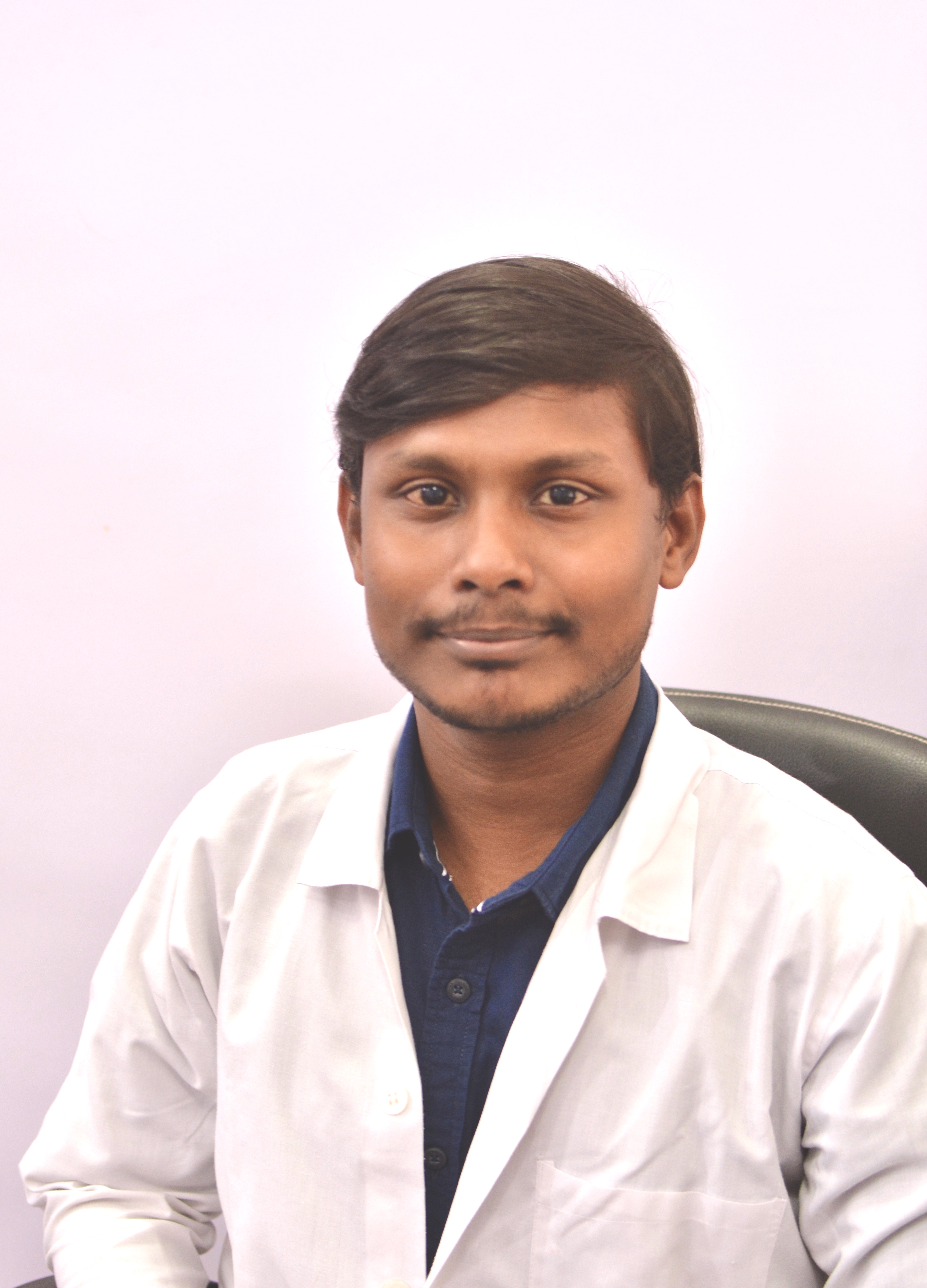
Fig. 1: Pre-operative smile.
Fig. 2: intra-oral view.
Treatment plan:
We decided on the amount of tooth movement based on the proclination, with our consultant orthodontist, and a Kesling-type set up was made, which was further waxed up to give us a fair idea of the final outcome. The final tooth positions post-orthodontic treatment were thus planned with the set-up and the treatment plan was presented to the patient. We discussed the plan including its pros and cons and for the restorative options for spaces, post-orthodontic treatment, were composite resin labial veneers or porcelain labial veneers. We discussed the importance of replacing missing lower right premolar and possible root coverage for the lower-left lateral incisor. The patient consented to treatment.
Treatment:
Phase one: Orthodontic treatment with metal brackets.
Phase two: Restorative treatment: Lithium disilicate veneers.
Orthodontic treatment concluded after 16 months.(fig. 3)
We analysed further, on occlusal analysis, the patient was noted to have a canine-guided occlusion on both sides on the lateral excursion. Periodontium of the teeth to be restored was fairly good. The upper lateral incisors lacked normal anatomic qualities, they were asymmetrical and seemed slender. The upper central incisors were medially inclined on axial inclination.
As part of treatment goals:
- we intended to close the gaps between upper anteriors.
- improve the tooth shape and symmetry
We discussed labial veneers in composite and porcelain materials. We informed the patient about the pros and cons of the two options. The patient was informed about the relatively faster rate of chipping, discolouration and fracture related to composite veneers. We discussed the maintenance related to composite veneers. Apart from the tooth preparation involved with porcelain veneers, we informed the patient about the limited scope of repair with porcelain.
The difference in cosmetic perceptions of composite veneers versus porcelain is not significant as per literature. The patient was, however, inclined to proceed with porcelain veneers, given the high success rates and acceptability. We made impressions of the upper and lower arches, and a facebow registration was done (fig. 5).
Fig. 3: Smile: post-orthodontic treatment.
Fig. 4: Intra-oral view, post-orthodontics.
Fig. 5: Facebow record.
The jaw relation, bite, was recorded in MIP, as we planned on a conformative approach with the restorations.
The mockup (fig. 6) was then assessed on a semi-adjustable articulator, including the lateral excursions (canine guidance).
We transferred the mockup to the patient’s existing dentition using a silicone index, loaded with bis-acrylic material after the teeth were spot etched. Post-mock-up transfer, the function was checked in static and dynamic occlusion. The excess was trimmed, and the mockup transfer was polished. We evaluated the smile line, phonetics and lip support, with photographs and videos (fig. 7). The patient was satisfied with the proposed final veneer designs.
Fig. 6: Mock up.
Fig. 7: Smile, post mockup transfer.
Shade selection was done using a DSLR camera and was evaluated for chroma, hue and value. We used an APT technique to prepare the teeth for veneers, where an aesthetic mock-up temporary is present on the teeth (proposed final outcome) to minimise the reduction required (fig. 9). We made silicone putty indices (fig. 8) to facilitate guided preparations for labial and incisal reduction. Depth cuts were made, to the desired dimensions.
Medium grit, followed by extra fine burs were used for the preparation and margin. A chamfer margin was selected. Preparations were then finished and polished (fig. 10, fig. 11). Impressions were made in a two-step, impression technique, using elastomeric putty and addition silicone light body impression materials.
Fig. 8: Use of silicone indices to guide tooth preparation.
Fig. 9: Depth cuts on APT.
Fig. 10: Final preparation.
Fig. 11: Close up of preparation
Literature supports a two-step technique, over a single step impression technique.
The provisional veneers were then re-fabricated using the silicone index, on the proposed mock-up, and placed on to the teeth after spot etching. The provisional restorations were finished and polished. Hygiene and post-care instructions were provided to the patient.
On the day of the cementation, the provisional restorations were removed using a spoon excavator, hand instrument. The preparations were then cleaned with a pumice slurry and rotary brush. The gingival health was assessed. The veneers were tried individually and then together, and assessed for marginal fit, proximal fit and overall aesthetics. A split-dam technique was used for rubber-dam isolation, for the anterior segment (fig. 12). The intaglio surfaces of the veneers were treated with 9.5% hydrofluoric acid, then rinsed thoroughly and dried with an air syringe (fig. 13). Silane was then applied and then the excess was blown off, with an air-syringe (fig. 14).
The preparations were etched with 37% phosphoric acid. Bonding agent was then applied to the preparations. Veneers were cemented, two at a time, starting from the midline and outwards, using light-cured resin cement (translucent shade). Excess was removed and the veneer margins were polished. The veneers were then checked for its function and final aesthetics were evaluated extra-orally (fig. 15).
Fig 12: Isolation
Fig 13: Etched veneers
Fig. 14: Silane application.
Fig 15: Post bonding of veneers
Discussion: The patient was satisfied with the outcome of the treatment. Smile makeovers rely heavily on the expectations of the patient. Aesthetics is subjective and a proper understanding of the patient’s requirements and expectations can help set the treatment objectives. As clinicians, we must assess these requirements and plan our treatment to incorporate the aesthetic expectations with function, in our treatment, accordingly.
Fig. 16: Post-operative smile.
Fig. 17: Smile, oblique
Fig. 18: After 15 month follow up.
The use of a semi-adjustable articulator enables us to assess the function of proposed designs before re-testing them in the patient with mock-up transfer. Mock-ups also allow patients to ‘test-drive’ their new smile and give them a clear understanding of what to expect from final restorations. The preparation is majorly in the enamel which enhances the adhesive properties of the veneers. This was possible with guided preparations and APT. Light cured resin cement used for bonding is known to have better colour stability, long term as per literature.
With regards to material selection, there has been no difference in terms of survival in the literature between lithium disilicate and feldspathic materials. Both materials provide excellent aesthetics. Composite veneers could definitely provide similar aesthetic outcomes; however, require a more intensive maintenance protocol and patient compliance. Digital planning and smile designing of this case and aligner technology for tooth movement, followed by veneers would have enabled better patient communication, and possibly more predictable outcomes.
Authors:
Dr Stephen Dsouza BDS | MSc (Aesthetic Dentistry) runs a private general practice in Mumbai, with a focus on Aesthetic and Restorative Dentistry. He practices minimally invasive dentistry.
Dr Shonali Patankar BDS | MDS (Orthodontics and Dentofacial Orthopedics) runs her private practice in Mumbai and practices orthodontics as well as general dental procedures.
In the realm of periodontal soft tissue surgery, a revolutionary wave is cresting: minimally invasive surgery. By promising precision and extraordinary ...
This article explores how artificial intelligence and augmented reality are revolutionising cosmetic dentistry—especially, how virtual smile previews help...
Guided Biofilm Therapy, or the GBT protocol, which is supported by scientific evidence, was developed in 2016 by EMS, together with academics, researchers, ...
Direct composite veneers are an excellent choice of treatment for aesthetic concerns. They are minimally invasive, have superior aesthetics, and are not as ...
Oral Submucous Fibrosis (OSMF) is a chronic and potentially malignant condition that affects millions of people worldwide, particularly in Southeast Asia. ...
This case report aims to demonstrate the decision-making and planning involved in treating a case of midline diastema and how an interdisciplinary approach ...
CZĘSTOCHOWA, Poland: Digital transformation in orthodontics is accelerating, and the diagnostic models generated by the orthodontic software DDP AI from ...
Smile Designing doesn’t always have to be about elaborate, extensive and invasive procedures but just sometimes routine procedures done with adherence to ...
BANGALORE, India: European dental manufacturers W&H Dentalwerk and Planmeca have joined forces on the dental market in India. Comprising a shared office...
Education
Live webinar Thu. 23 April 2026 8:30 pm IST (New Delhi)
Stanley M. Bergman’s retirement as CEO of Henry Schein marks the end of one of the most influential leadership tenures in modern dentistry and healthcare ...
TIRUCHIRAPPALLI, India: The integration of artificial intelligence (AI) into medical imaging can support diagnosis and reduce clinical workload. A new study...

 International / International
 Brazil / Brasil
 Canada / Canada
 Latin America / Latinoamérica
 USA / USA
 Austria / Österreich
 Bosnia and Herzegovina / Босна и Херцеговина
 Bulgaria / България
 Croatia / Hrvatska
 Czech Republic & Slovakia / Česká republika & Slovensko
 France / France
 Germany / Deutschland
 Greece / ΕΛΛΑΔΑ
 Hungary / Hungary
 Italy / Italia
 Netherlands / Nederland
 Nordic / Nordic
 Poland / Polska
 Portugal / Portugal
 Romania & Moldova / România & Moldova
 Slovenia / Slovenija
 Serbia & Montenegro / Србија и Црна Гора
 Spain / España
 Switzerland / Schweiz
 Turkey / Türkiye
 UK & Ireland / UK & Ireland
 China / 中国
 Pakistan / Pākistān
 Vietnam / Việt Nam
 ASEAN / ASEAN
 Israel / מְדִינַת יִשְׂרָאֵל
 Algeria, Morocco & Tunisia / الجزائر والمغرب وتونس
 Middle East / Middle East

 Dr. Hossam HamdyLive webinar
 Crystal SpringRegister now1CELive webinar
 Dr. Ana FerroLive webinar
 Dr. Renato Leonardo D.M.D.Register now1CE


To post a reply please login or register