State of the art dentistry is all about providing atraumatic and minimally invasive dental treatments to the patients, while improving the clinician's precision and efficiency. The first article of this 2-part series by Dr. Ridhima Uppal outlines the most essential concepts needed for beginners who are planning to incorporate magnification systems in their practice.
Upgrading our daily practice with a magnification system helps increase the clinician's precision in work as it not only provides a corrective and a magnified vision but also improves the ergonomics, thus increasing the work efficiency (Figure 1a & 1b).
Figure 1a: Improved ergonomics using magnifying loupes
Figure 1b: Improved ergonomics using Dental Operating Microscope
Two types of magnification systems (Figure 2) are in use-
a) Magnification loupes- most commonly used
b) Dental operating microscope.
Figure 2a: Magnification Loupes
Figure 2b: Dental Operating Microscope
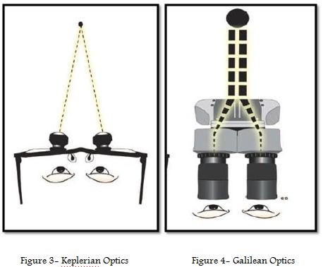
Magnification loupes work at a magnification of 2.5x-4x, where two monocular microscopes with side-by-side lenses are angled to focus on an object (Keplerian optics, Figure 3).
Being portable in nature, the loupes have an added advantage over a surgical operating microscope. But at the same time, loupes could result in eye fatigue as they cause converging of the eyes to view an image. The surgical microscope is however a complicated system of lenses that allows binocular viewing (Galilean optics, Figure 4) at a magnification of approximately 5x to 40x. Microscopes offer a powerful co-axial illumination with a higher magnification power over telescopic loupes.
Adapting to a magnification system is a steep learning curve and one should start with a pair of magnifying loupes thereupon strengthening the clinical skills.
Having an aided visual guidance gives an edge to enhance one’s fine motor skills that translate into minimal surgical trauma and smaller surgical field allowing better primary wound closure and consequently a rapid, comfortable, less painful, and non-inflammatory healing phase are some of the many advantages provided while using a magnification system.
The clinicians having been exposed to binoculars and cameras understand magnification as only a means of enhanced vision. But there is much more to what the eyes can see!
The perception of improved hand skills with the use of magnification has not yet been appreciated. The improved ergonomics benefit the clinician in avoiding musculoskeletal problems and reduced eye fatigue. Adding to all these benefits, a magnification system is a strong tool for patient education and motivation and for self-evaluation of one's own work thus helping in improving the quality of day-to-day practice.
'' I can do better; if I can see better; but I can do the best if I can see even better''
Today magnification has revolutionized dentistry all around the world. Higher magnification with a dental operating microscope (DOM) is clearly an asset, ...
This article series is designed to help dental students and professionals navigate the path to an international dental career with clarity and confidence. ...
In Part 1 of this series, we examined how dental students can begin preparing early to establish a global-ready profile. In Part 2, we focus on interns, ...
COVID-19 is having an unprecedented impact on the global economy, including that of the dental industry. Most dental clinics are shut and a few are offering...
Dr Prajwalit Kende and his team, Dept of Oral & Maxillofacial Surgery, Govt Dental College & Hospital, Mumbai has innovated "Z-plate" for the management of ...
COVID-19 is having an unprecedented impact on the global economy, including that of the dental industry. Most dental clinics are shut and a few are offering...
COVID-19 is having an unprecedented impact on the global economy, including that of the dental industry. Most dental clinics are shut and a few are offering...
COVID-19 is having an unprecedented impact on the global economy, including that of the dental industry. Most dental clinics are shut and a few are offering...
Education
Live webinar Mon. 13 July 2026 9:00 pm IST (New Delhi)
MUMBAI, India: Can clinicians identify patients at higher risk of implant complications before surgery? Can understanding the patient’s immune response ...
Implant dentistry has evolved significantly through advances in implant design, surface technology and digital workflows. However, the process of osteotomy ...
While implant dentistry often focuses on implant design, surface characteristics and surgical protocols, the role of drill material in osteotomy preparation...

 International / International
 Brazil / Brasil
 Canada / Canada
 Latin America / Latinoamérica
 USA / USA
 Austria / Österreich
 Bosnia and Herzegovina / Босна и Херцеговина
 Bulgaria / България
 Croatia / Hrvatska
 Czech Republic & Slovakia / Česká republika & Slovensko
 France / France
 Germany / Deutschland
 Greece / ΕΛΛΑΔΑ
 Hungary / Hungary
 Italy / Italia
 Netherlands / Nederland
 Nordic / Nordic
 Poland / Polska
 Portugal / Portugal
 Romania & Moldova / România & Moldova
 Slovenia / Slovenija
 Serbia & Montenegro / Србија и Црна Гора
 Spain / España
 Switzerland / Schweiz
 Turkey / Türkiye
 UK & Ireland / UK & Ireland
 China / 中国
 Pakistan / Pākistān
 Vietnam / Việt Nam
 ASEAN / ASEAN
 Israel / מְדִינַת יִשְׂרָאֵל
 Algeria, Morocco & Tunisia / الجزائر والمغرب وتونس
 Middle East / Middle East

 Dr. Fernando FranchLive webinar
 Dr. Nicolas OuelletRegister now1CELive webinar

 Dr. Nisha D’Silva BDS, MSD, PhD, Dr. Kıvanç Bektaş-KayhanRegister now1CELive webinar
 Federico ZunicaRegister now1CE


 - Dr. Ridhima Uppal")


- Dr. Ridhima Uppal")


")


")
")
")


To post a reply please login or register